Consultation
Macular Hole
Eye Conditions
Cataract
CSCR
Diabetic Retinopathy
Epiretinal Membrane
Flashes and Floaters
Glaucoma
Macular Degeneration
Macular Hole
Retinal Detachment
Retinal Vein Occlusion
Uveitis
Vitreomacular Traction
Investigations
Procedures
Macular hole is the formation of a round gap at the centre of the macula. This leads to blurring of vision and distortion of images. A blind spot may appear at the middle of vision.


MacularHole
OCT Scan
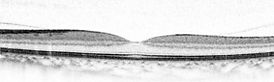
Cross section view of normal macula
Leads to

Cross section showing thickened and
distorted macula with a full-thickness defect at the centre
Blurring and distortion of images
Macular hole is treated by vitrectomy surgery. The surgery is performed under local anaesthesia with minimal discomfort. With modern techniques, sutures are usually not required. A bubble of gas is injected to keep the hole dry and allow it to heal. It is helpful to look down for 3 to 5 days to keep the macular hole above fluid. You must not travel by air while gas bubble remains.

Vitreous is removed through tiny cuts on the sclera (white of eye) using an automatic cutter.

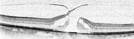
Macular Hole

Gas Bubble
A thin membrane on the
surface of the macula is stained with a dye and peeled with forceps to release tension from the hole.
Gas bubble is injected to keep the hole dry.
What is a macular hole?
A macular hole is a round defect in the central part of the macula. It causes blurring and distortion of the central vision, which may also develop a blind spot. Without treatment, macular holes progressively enlarge and most patients end up with a legally blind eye (less than 6/60). Because the peripheral retina is not affected, macular holes do not lead to total blindness.
Who are affected?
Macular holes most commonly occur in people aged in their 60s and 70s. Women are affected slightly more often than men. Uncommonly, macular holes can occur in younger people as a complication of retinal injuries, swelling or surgery.
What causes a macular hole?
The cavity inside the eye is filled with a gel-like substance called the vitreous. In youth, the vitreous is stuck to the surface of the retina. With aging, the vitreous shrinks and separates away from the retina. Sometimes the vitreous adheres tightly to the macula and cannot separate easily. The persistent pull of the vitreous on the macula causes blisters to form, which break down to develop into a hole.
How is a macular hole diagnosed?
Although macular holes can be seen through the examination microscope, OCT scan has become the standard for its diagnosis and evaluation. OCT scan uses computer-controlled laser beams to build cross-sectional images of the macula, offering unprecedented details of its structure. Fluorescein angiography may be performed to assess the retinal circulation and to exclude possible underlying causes of macular hole.
When should a macular hole be treated?
Macular holes do not need to be treated urgently, but earlier surgery offers a better outcome. Long-standing macular holes may not be treatable.
How is a macular hole treated?
Macular holes are treated by a technique called vitrectomy. Modern vitrectomy technique uses tiny wounds and do not require sutures. Three small wounds are made on the white part of the eye, through which an infusion tube, light source and vitrectomy cutter are inserted. After the removal of the vitreous gel, the surface of the macula is stained with a dye. A thin layer of tissue on the macula is removed using a pair of fine forceps to relieve tension from the hole. A bubble of gas is injected into the eye cavity to keep the hole dry and allow it to heal. Patients are required to look down as much as possible for 3 to 5 days to keep the gas bubble placed on the hole.
What are the benefits and risks of having surgery for a macular hole?
The aim of the surgery is to close the hole and improve vision. Over 90 % of macular holes are expected to close after a single attempt, with the vision improving in the majority of the patients. The success rate is lower with long-standing holes. It is usually not possible to achieve a completely normal vision.
Overall risk of complication is low, but can include the following:
More common: Cataract, failure to close hole
Less common: Retinal detachment, high eye pressure, temporary bleeding
Rare: Infection, severe bleeding, unexplained poor vision, recurrent macular hole
Cataract can develop months or years after the vitrectomy and may require surgery. Cataract refers to the lens inside the eye becoming hazy. Modern surgical techniques have made cataract surgery safe and efficient. The delicate retina can be injured during vitrectomy and result in a retinal tear or detachment. Retinal tears found during the surgery will be treated immediately, but if the retina becomes detached after the surgery, a second procedure will be needed to repair it. As for all eye surgeries, bleeding and infection are rare possibilities despite efforts to minimise their risk. Recurrence of the hole is rare but possible.