Retinal Vein Occlusion
Eye Conditions
Cataract
CSCR
Diabetic Retinopathy
Epiretinal Membrane
Flashes and Floaters
Glaucoma
Macular Degeneration
Macular Hole
Retinal Detachment
Retinal Vein Occlusion
Uveitis
Vitreomacular Traction
Investigations
Procedures
Retinal vein occlusion (RVO) is blockage of a vein draining the retina. Blockage of the central vein within the optic nerve leads to Central RVO, affecting all of the retina. Blockage involving a smaller vein leads to branch RVO, which affects a portion of the retina.

Branch RVO

Central RVO
The blockage in the vein reduces blood flow, limiting oxygenation and nutrient supply to the affected part of the retina. The pressure in the vein also rises. This leads to dilation of the veins and leakage of serum and blood, causing retinal swelling and haemorrhage. Ultimately, the affected part of the retina can become irreversibly damaged by the lack of oxygen.

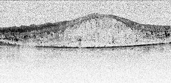
OCT scan showing macular swelling. Fluorescein angiography (right) shows damage to blood vessels.

Treatment of Retinal Vein Occlusion
Laser therapy is used to treat macular swelling and to prevent complications of ischaemia (damage due to lack of blood flow).

Macular oedema

Intraocular injections have become the main treatment of macular oedema.
Anti-VEGF agents are most commonly used, but steroid injections are also effective.


Vitrectomy may be required for persistent vitreous haemorrhage or retinal scarring and detachment.

Before Surgery
After Surgery
