Consultation
Central Serous Chorioretinopathy
Eye Conditions
Cataract
CSCR
Diabetic Retinopathy
Epiretinal Membrane
Flashes and Floaters
Glaucoma
Macular Degeneration
Macular Hole
Retinal Detachment
Retinal Vein Occlusion
Uveitis
Vitreomacular Traction
Investigations
Procedures
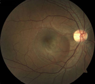
Leakage of fluid through a defect in the retinal pigment epithelium leads to build up of fluid under the macula. The cause is unknown in most cases but psychological stress, taking steroids, pregnancy and conditions such as Cushing syndrome are known risks.
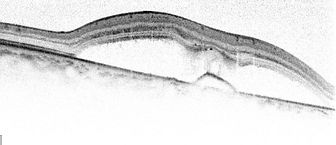
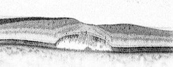
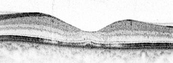
OCT scan showing build up of fluid under the macula and disturbance of retinal pigment epithelium
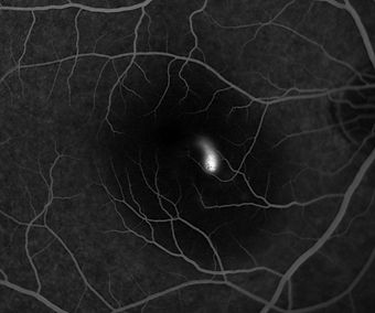
Fluorescein Angiogram
Diagnosis is made through clinical examination, aided by OCT scan and fluorescein angiography. Most cases resolve without treatment after 3 months, but subtle visual changes can remain permanently. In around 15 % of cases, CSCR proves to be chronic or recurrent, with a significant risk of visual impairment.
Management of CSCR
Most patients are followed without treatment because spontaneous recovery is expected. Steroid treatment (e.g., oral prednisolone, steroid skin ointment) should be ceased if possible. Life stresses should be controlled. Regular exercise and adequate sleep are advised. Should the fluid fails resolve by around 3 months, intervention will be considered because permanent visual impairment becomes likely.
If fluorescein angiography shows focal leakage well away from the centre of the macula, it can be treated with gentle laser therapy. When the leakage is widespread, or is located close to the central macula, photodynamic therapy can be considered. This involves an injection of verteporfin into the blood stream and treating the macula with a low-power laser to activate the drug and heal the leaking spots. Oral medications have been tried with unproven efficacy.

Rarely, chronic CSCR can be complicated by the development of abnormal blood vessels in the choroid. Treatment with intraocular injection of anti-VEGF agent is essential to avoid severe vision loss.
In central serous chorioretinopathy (CSCR), fluid accumulates under the macula causing a round or oval blister. Symptoms include blurring of vision, distortion, discoloured patch in central vision or images appearing smaller.