Consultation
CARE
OFFERED
Investigation
Advances in medical technology have dramatically changed the way the patients are cared for. Modern instruments allow examination of your eyes in unprecedented detail and precision. The information gathered from these investigations add clues to diagnosing your eye condition and to monitor its progress.
Digital photography
Photography of the retina has been an important tool for managing retinal disorders for almost a hundred years. Along with the sketches made by the doctor, retinal photographs allow an accurate recording of the disease process and treatment responses. The advent of digital photography and computerised archiving have made this important tool more accessible and useful than ever.
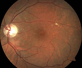
Optomap imaging

Traditional optical cameras have a limited field of view. In other words, it only shows a small area of the retina at a time. Optomap imaging offers an unparalleled view of your retina by using scanning lasers to obtain images. The resulting photograph shows a very wide field of view, allowing your entire retina to be examined at once. This technology can also be used for fluorescein angiography, complementing conventional angiography with the benefit of a more complete peripheral coverage.
Optical coherence tomography (OCT)
OCT scan has revolutionised diagnosis and treatment of retinal disorders and has become an essential part of the modern retinal practice. It is a quick and non-invasive test that allows the layers of the retina and underlying tissues to be assessed in microscopic details. OCT uses a sweeping beam of laser to scan an area of the retina. The optical properties of various parts of the retina are computed to build two and three-dimensional images of the retina.

Fluorescein and ICG Angiography
Angiography is a diagnostic procedure that studies blood vessels and circulation. A dye is injected into the bloodstream to outline the blood vessels and a special camera is used to record the blood flow. Retinal angiography is commonly performed using a fluorescein dye, but an ICG dye may be used to study the deeper tissues.
Fluorescein and ICG angiography is much safer than heart or brain angiography. Nevertheless, it is an invasive procedure that requires injection of a dye into your vein. It can cause a brief episode of nausea and rarely, some patients will vomit. The dye colours your skin complexion for a day until it is passed through your urine. Anytime a drug or chemical is injected into the blood stream, there is a small risk of severe allergic reaction. This is why angiography can only be performed in a medical practice. Severe side effects are extremely uncommon and the information gathered can be of critical importance to managing your eye condition.

Visual field test
While the vision chart tests your central vision, it does not provide information about the peripheral vision. The visual field test maps the sensitivity of the overall field of vision by projecting targets in your peripheral vision. The visual field test is most commonly performed for glaucoma but it is also valuable for assessing disorders of the retina, optic nerve and brain.

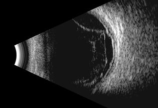
Ocular ultrasound scan
Ultrasound scans use sound waves to build an image of the eye and allow measurements of its internal structures. It is entirely harmless and pain-free. Ultrasonography is extremely useful when the view of the internal structures are obscured, for example, blood.
External investigations
Further investigations may be necessary for complex conditions.
These may include:
• Blood tests
• CT, MRI and body ultrasound scans
• Electrophysiological tests